Abstract
Objective
Tomography is the gold standart method for gunshot injury cases but there are limited information in the literature on the use of tomography in shooting analysis. This study aimed to examine the findings required for shooting reconstruction, clinical, forensic evaluation and to show the advantages-disadvantages in gunshot injuries.
Material and Methods
The head of 12 Adana sheep and Canik Mete TP9 9x19 mm were used. Shots were made to the frontal, temporal, and occipital regions from distances of 50 cm, 1, 5 and 10 meters. Vimago GT30 (Epica animal health, USA) computed tomography was used for scanning.
Results
The time required to obtain images for all heads were less than 1 minute. Entry and exit wounds findings were obtained from axial and sagittal images for frontal and occipital shots, and from axial and coronal images for temporal shots. Bullet core trajectory and brain damage were examined from axial and sagittal images for frontal and occipital shots, from axial and coronal images for temporal shots, and Hounsfield unit (HU) values were measured from axial images for all shots. HU values -808±164 (from -530 to -1024) in cavitation area, +2512±200 (from +2332 to +3071) in pathological tissues, +831±85 (from +707 to +966) in brain tissue, and +2683±359 (from +2128 to +3071) in bone were found.
Conclusion
Tomography provides critical information about the location and size of entry-exit wounds, HU values in different tissues, trajectory, cavitation and intracranial foregin bodies. It is possible to determine the azimuth and vertical angles necessary for shooting reconstruction using tomography.
Keywords: Bullet core trajectory, cranial gunshot injuries, computed tomography, shooting reconstruction
INTRODUCTION
Cranial gunshot injuries (GSIs) are severe traumas associated with high mortality and morbidity. Computed tomography (CT) is the gold standard imaging modality for these cases (-). Cranial imaging is recommended as soon as possible after arrival at the hospital. Brain damage depends on the type of firearm, the distance of the shot, and the entry angle, mass, and velocity of the bullet. While the majority GSI cases die at the scene, mortality can be reduced in those who reach the hospital with rapid, appropriate and aggressive treatment (, ). Tomography provides critical information about the location and size of entry and exit wounds, determination of brain damage, bullet trajectory, cranial bone fractures, detection of foreign bodies, and bullet residues (bullet core or jacket fragments). It plays a pivotal role in surgical decision-making and in determining the surgical strategy. Given the high mortality rate associated with GSIs, physicians must make rapid decisions. At this stage, CT provides rapid information among all radiological imaging methods (, , , ).
The first postmortem CT scan was reported in the late 1970s in a fatal cranial GSI. The VIRTOPSY project, presented in 2003, described minimally invasive imaging methods to complement traditional autopsies. Medical imaging methods, particularly CT, are increasingly used for forensic purposes (-). Images obtained using this non-invasive method can be re-examined, stored, shared, presented in courts, and used to create 2D and 3D images (-). CT has proven particularly effective in detecting fractures, foreign bodies, and gases (-). Forensic physicians detect entry-exit wounds and trajectory, determine the type of weapon, and ascertain the cause of death (-). Rapid and accurate identification of victims is essential after disasters. Interpol has deemed CT-based data suitable and useful. The interpretation of reconstructed images, independent documentation from observers, the ability to reconstruct various images long after the event, the sharing of workload through digital transmission of CT data, and the reduction in time and expertise required in the disaster area have demonstrated that it is a valuable tool for disaster victim identification. Age determination and identification through comparison of antemortem and postmortem CT images have become possible with CT (, ).
Limited literature exists on the use of CT for crime-scene and shooting reconstruction. CT scans detect bullet fragments in decomposed, skeletal, mummified, or charred corpses. Limited data regarding ammunition and caliber support the usefulness of CT. CT findings are valuable in cases with minimal skin findings or intraoral GSIs. While limited number of publications report that shooting distance with CT, these studies are generally based on animal experiments and the detection of gunshot residue (-). Shooting reconstruction is the study conducted to identify the person who pulled the trigger and the shooting location (). Azimuth (horizontal) and vertical angles are the necessary for shooting reconstruction (). Existing literature provides insufficient information on the contribution of CT to shooting reconstruction by determining azimuth and vertical angles. This study aimed to fill this gap. We aimed to examine the findings required for shooting reconstruction and discuss the advantages and disadvantages of CT in GSIs.
MATERIALS and METHODS
Study Population
Although no ideal subject species has been defined in the literature for use in GSIs, organ models, pigs, and sheep have been used most frequently in ballistic studies (-). Sheep were preferred for the study because pigs were unavailable after the regional earthquake. Moreover, sheep were more accessible and required fewer anaesthetic agents for control purposes. The study utilized the heads of 12 Adana breed sheep. The heads were obtained at no cost from a slaughterhouse that processes sheep daily, and the animals were automatically slaughtered at the distal part of the C1 vertebra (Atlas). External examination revealed no differences among the heads used. The heads were stored at +4 °C in sterile conditions and transported to the experimental area.
Ammunition used were 9×19 mm calibre M822 type (Full Metal Jacket) rounds (340 m/s), fired from a Canik Mete TP9 semi-auto handgun; this calibre is the most common in civilian injuries. Experienced tests were performed by the staff of the xxxSpecial Operations Directorate, Special Operations Department, General Directorate of Security, Ministry of Internal Affairs, at the same department shooting range.
Test Shots Plan
Test shots were performed from distances of 0.5, 1, 5, and 10 meters, targeting the frontal, temporal and occipital regions ().
Table 1. Fire region and distance of heads
Head | - | Region | - | - | Distance | ||
|---|---|---|---|---|---|---|---|
FR | T | OC | 0.5 meter | 1 meter | 5 meter | 10 meter | |
1 | X | - | - | X | - | - | - |
2 | - | X | - | X | - | - | - |
3 | - | - | X | X | - | - | - |
4 | X | - | - | - | X | - | - |
5 | - | X | - | - | X | - | - |
6 | - | - | X | - | X | - | - |
7 | X | - | - | - | - | X | - |
8 | - | X | - | - | - | X | - |
9 | - | - | X | - | - | X | - |
10 | X | - | - | - | - | - | X |
11 | - | X | - | - | - | - | X |
12 | - | - | X | - | - | - | X |
FR: Frontal, T: Temporal, OC: Occipital
Cranial Imaging
Heads were positioned with the nape and chin aligned, keeping the heads in the midline. Axial images were obtained using a Vimago GT30 (Epica Animal Health, USA) high-resolution CT scanner. Tomography parameters were as follows: 120 kVp, 80 mAs, gantry rotation time =0.6 s, slice thickness =0.35 mm. The time required for imaging was recorded, and a veterinarian appropriately disposed of the imaged heads.
CT images were evaluated by two authors. The axial images and the sagittal and coronal reformats, recorded in DICOM format, were transferred to digital storage and analysed by researchers on personal computers. During the evaluation, factors such as the location, diameter, and angle of the entry wound, the presence of bullet core residue, the bullet trajectory, brain tissue damage, bone fractures, the distribution of bone fragments, Hounsfield unit (HU) values, and the location and diameter of the exit wounds were examined.
Ethical approval for the study was received from the Çukurova University Experimental Animals Local Ethics Committee on 30.04.2024 (meeting number: 4, decision number: 6).
Statistical Analyses
For this descriptive study, HU values were performed using SPSS 20.0 (Chicago, IL), findings are presented as mean ± standard deviation (minimum-maximum).
RESULTS
The time required to obtain images for all 12 heads was less than 1 minute. Findings of entry and exit wounds were obtained from axial and sagittal images for frontal and occipital shots and from axial and coronal images for temporal shots. Bullet trajectory and brain damage were examined on axial and sagittal images for frontal and occipital shots, on axial and coronal images for temporal shots; HU values were measured on axial images for all shots. HU values -808±164 (from -530 to -1024) in cavitation area, +2512±200 (from +2332 to +3071) in pathological tissues, +831±85 (from +707 to +966) in brain tissue, and +2683±359 (from +2128 to +3071) in bone were found ( and ).
Table 2. Cranial computed tomography findings of entry and exit wounds
Head | Entry wounds angle | Entry wounds region | Enrty wounds defect region | Enrty wounds diameter | Exit wounds region | Exit wounds defect region | Exit wounds diameter |
|---|---|---|---|---|---|---|---|
1 | 9.75° Azimuth -31.7°Vertical | FR region right of midline | Skin Bone | 1.7x3.8 mm 9.4x88.0 mm | OC bone, right arcus of C1 | Bone | 150x270 mm |
2 | 0° Azimuth -3.5° Vertical | Right T bone | Skin | 3.3x3.0 mm | Left T bone | Bone | 20.0x14.0 mm |
3 | 1.0° Azimuth -20.0° Vertical | OC region midline | Bone | 9.2x12.0 mm | Nose, right of midline | Bone | 3.3x5.0 mm |
4 | 6.0° Azimuth -23.0° Vertical | FR region left of midline | Skin Bone | 3.0x3.8 mm 7.5x16.0 mm | OC bone, Midline | Bone | 3.8x3.5 mm |
5 | 14.0°Azimuth -8.0° Vertical | Right T bone | Skin | 4.0x5.2 mm | Left ear | Skin | 7.0x6.0 mm |
6 | 10.0° Azimuth -4.0° Vertical | OC region Midline | Bone | 28.0x23.0 mm | Maxilla bone | Bone | 7.4x12.0 mm |
7 | 6.6° Azimuth -23.0° Vertical | Vertex-FR region right of midline | Bone | 5.8x8.0 mm | OC bone right of midline | Bone | 7.2x4.7 mm |
8 | 12.1° Azimuth -6.3° Vertical | Right T bone | Skin | Milimetric | Left T bone | Skin | Milimetric |
9 | 0° Azimuth -20.0° Vertical | OC region midline | - | 8.0x13.0 mm | Nasal region right of midline | - | 4.1x5.2 mm |
10 | 10.0° Azimuth -12.7° Vertical | Maxilla region right of midline | Skin Bone | 6.1x6.5 mm 10.0x12.0 mm | Right arcus of C1 | Bone | 4.0x10.0 mm |
11 | 8.0° Azimuth 1.2° Vertical | Right T bone | - | 7.1x5.3 mm | Left T bone | Skin | 6.6x5.7 mm |
12 | 28.0° Azimuth 21.0° Vertical | Posterior arcus of C1 | Bone | 10.0x14.0 mm | Nasal bone Midline | Bone | 10.0x10.0 mm |
FR: Frontal, OC: Occipital, T: Temporal, C1: First cervical vertebra
Table 3. Cranial computed tomography findings of bullet core trajectory and brain damage
Head | Bullet core trajectory | Location | HU | Brain damage |
|---|---|---|---|---|
1 | At the posterolateral line in the right lobe From cranial to caudal | Cavitaion Pathological Brain Bone | -530 2351 889 2503 | Fragmentation in FR sinusoids Fragmented bone pieces and cavitattion in the FR and OC lobes OC and C1 vertebral bone damage |
2 | From right to left At the midline From cranial to caudal | Cavitaion Pathological Brain Bone | -1024 2439 824 2388 | Fragmentation in T bone Fragmented bone pieces and cavitation in the T lobe |
3 | In the midline From cranial to caudal From posterior to anterior | Cavitaion Pathological Brain Bone | -641 2578 837 2230 | Large cavitation in OC lobe Fragmentation in maxillary sinusoids Defect In the nasal bone and distal nose skin |
4 | At the posterolateral line in the right lobe From cranial to caudal | Cavitaion Pathological Brain Bone | -886 2441 854 3071 | Fragmentation in FR sinusoids Fragmented bone pieces in the FR and OC lobes OC bone damage |
5 | From right to left From cranial to caudal | Cavitaion Pathological Brain Bone | -1024 2659 715 3071 | Fragmentation in T bone Fragmented bone pieces and cavitation in the T lobe |
6 | At the anterolateral line From cranial to caudal | Cavitaion Pathological Brain Bone | -665 2572 898 2601 | Fragmentation in FR and maxiller sinosioids Fragmanted bone pieces and cavitattion in the FR and OC lobes |
7 | At the posterolateral line in the right lobe From cranial to caudal | Cavitaion Pathological Brain Bone | -708 3071 966 3071 | Wide fracture line starting from entry wounds in the FR bone and extending posteroleteral line Fragmanted bone pieces and cavitattion in the FR and OC lobes |
8 | From right to left At the midline From cranial to caudal | Cavitaion Pathological Brain Bone | -816 2594 899 3071 | Fragmentation in T bone Fragmented bone pieces and cavitation in the T lobe |
9 | At the midline From cranial to caudal From posterior to anteior | Cavitaion Pathological Brain Bone | -801 2361 825 2766 | Cavitation in OC lobe Fragmentation in maxillary sinusoids and midline bones |
10 | At the posterolateral line in the right lobe From cranial to caudal | Cavitaion Pathological Brain Bone | -993 2452 707 2128 | Extensive tissue loss along the maxillary sinosoids, nasopharyngeal region and posterior jaw |
11 | From right to left From caudal to cranial | Cavitaion Pathological Brain Bone | -770 2583 752 2513 | Fragmentation in mandible, orbital wall Cavitation in brain tissue |
12 | At the anteromedial line From caudal to cranial | Cavitaion Pathological Brain Bone | -978 2332 713 3071 | Bullet core passed from the left to the right of the midline in the nasopharyngeal region, creating fragmentation in the bones and extensive cavitation in the tissue |
FR: Frontal, OC: Occipital, T: Temporal, C1: First cervical vertebra, HU: Hounsfield unit
DISCUSSION
In recent years, advancements in tomography and computer technology have created new opportunities for radiologists, clinicians, surgeons, and forensic physicians to evaluate GSI cases. Research on the advantages and disadvantages of CT in forensic cases, particularly in cranial GSIs, has demonstrated that CT is a suitable and beneficial method () (, ).
Table 4. Advantages and disadvantages of computed tomography
Advantages Entry wounds localization can be detected Diameter of entry wounds can be measured Two angles of entry wounds can be calculated Skin and bone defect can be detected Bullet core residue can be detected Fargmented bullet core can be detected Bullet core trajectory can be detecetd Intracerebral, intraventricular, epidural, subdural, subarachnoid hemorrhage can de shown Cerebral edema, pneumocephalus, shift, herniation can be detected Bone fragmantation can be detected Distrubution of fragmented bone pieces can be shown Soft tissue damage, cavitation can be shown Presence and distribution of foreign objects can be detected Hounsfield unit can be calculated Entry and exit wounds can be differentiated Exit wound localization can be detected Diameter of exit wounds can be measured Cause of death can be determinated It is a non-invasive method Body integrity is perserved Examination can be repeated with the same or different method Data can be stored digitally Data can be re-evaluated and commented on by different people and platforms Segmentation and seperation of tissues provide visual dissection in different planes Provides time and speed advantage Risk of infection transmission is low Contributes to the crime scene, wound and shooting reconstruction Allows 3Dimension reconstruction | Disadvantages May not provide information in sudden deaths Necessity of device, computer, HIMS, DICOM, PACS Cost Artifacts Radiation risk Experience required in technology, computers, radiology |
HIMS: Hospital information management system, DICOM: Digital ımaging and communications in medicine, PACS: Picture archiving and communication systems
Multi-slice CT scanners equipped with 64 detectors rotate around the patient at three revolutions per second. At this speed, whole-body screening takes less than 10 minutes, offering significant dose-time advantages (, ). In our study, the time to obtain images with the multi-slice CT device designed for veterinary use was less than 1 minute in all cases, highlighting CT’s efficiency.
During the evaluation, the diameters of skin and/or bone defects were measured after their locations were determined. In frontal and occipital shots, the skin entry wounds were smaller than the bullet diameter, while the bone entry wounds were equal to or larger than the bullet diameter. Millimetric entry wounds were observed on the skin in shots to the temporal region. The skin elastic structure resists penetration, causing the tissue to pit and stretch until the elasticity limit is exceeded, after which the bullet pierces the skin (, ). This explains why the observed skin entry wound was smaller than the bullet diameter in this study.
The azimuth angle was determined by measuring the angle formed between the entry wound and the midline in axial images. In contrast, the vertical angle was measured as the angle between the entry wound and the midline on sagittal and coronal images. The vertical angle represents the upward (+) or downward (-) component of the bullet’s trajectory, while the azimuth angle reflects the lateral direction as seen from above. For forensic shooting analysis and reconstruction, knowing the impact angles and entry wound characteristics is crucial (). Our most important findings suggest that CT provides essential data for forensic shooting analysis (shooting reconstruction studies).
Our evaluations also revealed that bullets create varying defects in the skin and bone. Two factors contribute to the variable sizes of exit wounds in GSIs. First is the bullet tumbling and exiting the body at different angles, and the second is the fragmentation and deformation of the bullet (, ). The CT findings in our study aligned with established principles of wound ballistics.
Cranial CT HU values help differentiate tissues: -1000 represents air, negative values near zero (-10, -20) indicate fatty tissues, positive values near zero (+10, +30) indicate fluid structures, +300 to +350 indicate calcified tissues, and +1000 represents bone, the densest structure (). Advanced reconstruction algorithms enable more detailed measurement of tissue attenuation and provide a wider HU range. The application used in this study had an HU range of -1024 to +3071. Air was represented by -1024 and bone by +3071. Bullet core residue, depending on its copper, lead, aluminium, or zinc content, can appear with extremely high HU values, such as +10,000 (, ). In this study, HU measurements showed that air largely replaced brain tissue in the cavitation area, while bone fragments were detected as pathological tissue in the brain. No bullet core residue was found in brain tissue. The absence of bleeding from the vessels was attributed to insufficient blood flow in the cranial region following decapitation.
The entry wound, bullet trajectory, exit wound, and HU values of subject 10 (impacting the frontal region from a distance of 10 meters) are presented as examples in and .
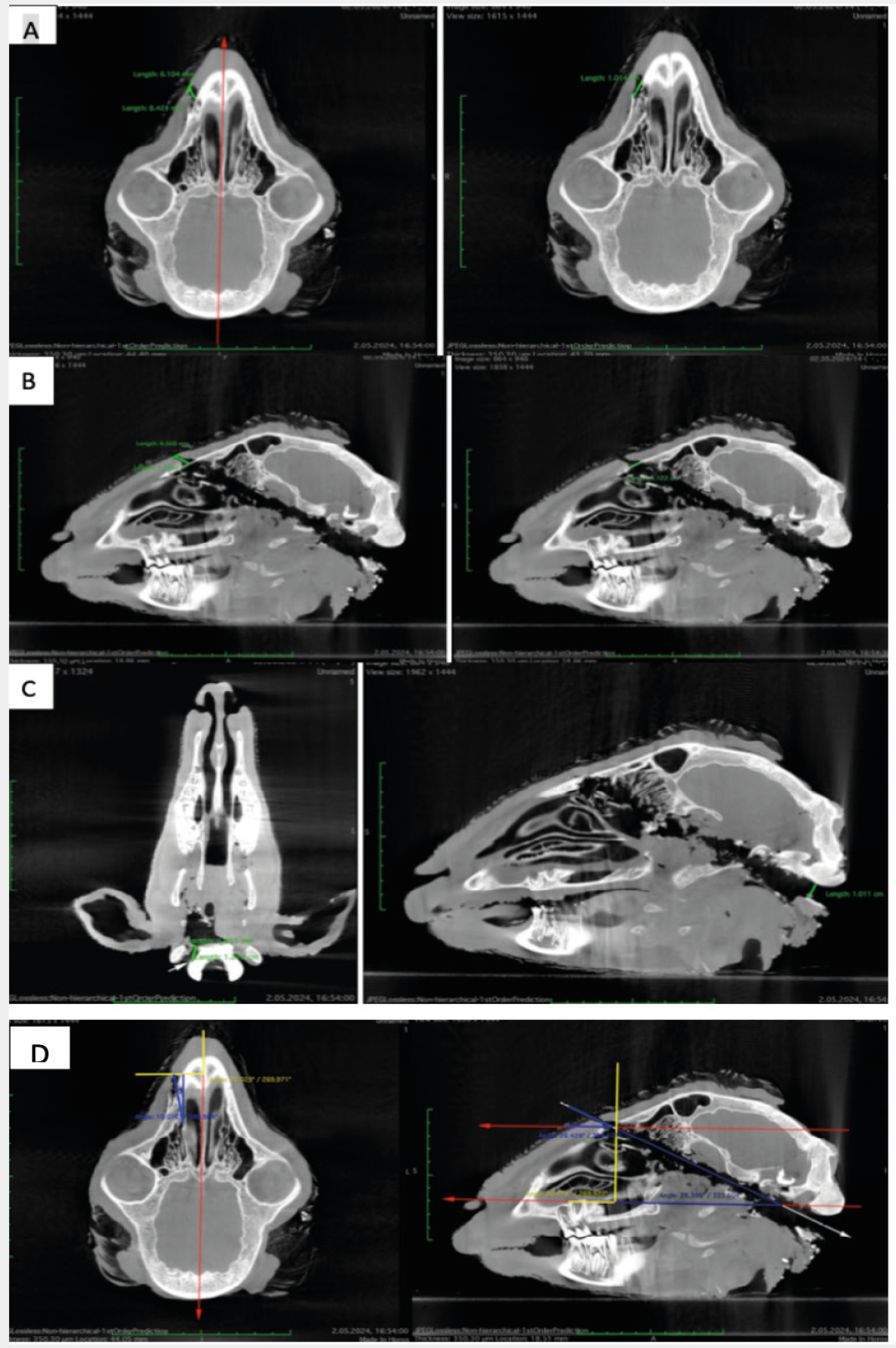
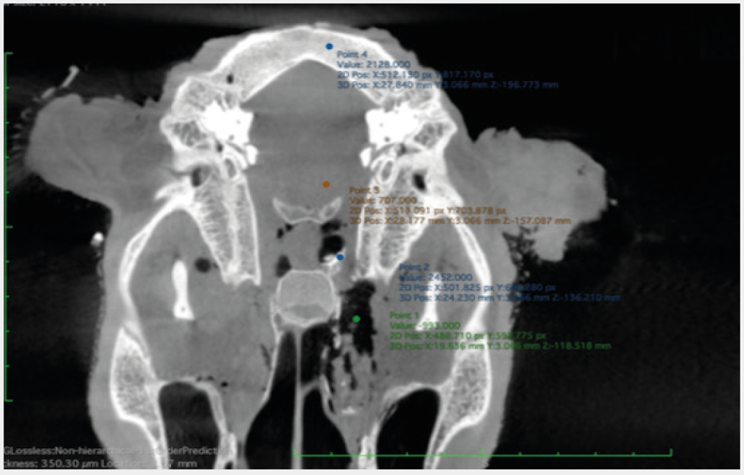
HU: Hounsfield unit
While the need for infrastructure such as hospital information management system, digital imaging and communications in medicine and picture archiving and communication system can be seen as a disadvantage, the “Regulation on Health Information Management Systems (HIMS)” dated August 22, 2022, indicates that the current health system in our country has the necessary infrastructure to support the evaluation of cranial GSI cases using CT (). During the study, the ability to share, export, and use DICOM-formatted data on personal computers enabled physicians to interpret images independently. Similarly, the HIMS regulation enables physicians to access and report data in certain situations, and telemedicine-integrated systems allow remote physicians to report data rapidly and without interruption. Another disadvantage is the presence of artifacts, including linear and ring artifacts, partial-volume effects, noise and motion artifacts, windmill effects, and beam hardening. However, these artifacts can be mitigated through various techniques and imaging adjustments ().
Study Limitations
The limitations of this study include its animal-based design and the small sample size. The anatomy of the sheep head is similar to that of humans, and the small number of subjects does not alter the findings. The study demonstrated that, regardless of shooting distance and location, CT provides the necessary information for shooting reconstruction. The second limitation was that we could not observe intracranial haemorrhage because of insufficient blood volume resulting from post-decapitation bleeding. However, since CT has been shown in previous studies to be a suitable method for demonstrating intracranial haemorrhage, this will not negatively affect the study’s value.
CONCLUSION
Cranial CT has numerous advantages, including speed and efficiency. Tomography provides critical information on the location and size of entry and exit wounds, the azimuth and vertical angles of entry, HU values in different tissues, the trajectory of the bullet core, cavitation, and bone fragmentation, and the distribution of bone fragments. The most important result of this study is that we have demonstrated that CT is a fast, non-invasive, and valuable method for forensic ballistic examination and forensic shooting analysis
Footnotes
Ethics
Ethics Committee Approval: Ethical approval for the study was received from the Çukurova University Experimental Animals Local Ethics Committee on 30.04.2024 (meeting number: 4, decision number: 6).
Informed Consent: It is not necessary for animal studies.
Acknowledgments
We would like to express our gratitude to Turkish Ministry of Internal Affairs, General Directorate of Security, Special Operations Department workers for their efforts.
This study was presented as oral presantation at 19th International Forensic Medicine Congress, 23-29 September 2024, Antalya, Türkiye.
Authorship Contributions
Concept: İ.A., G.İ.Ö., Design: İ.A., G.İ.Ö., A.S.K., Data Collection or Processing: İ.A., G.İ.Ö., A.S.K., K.İ., Ç.A., Analysis or Interpretation: İ.A., G.İ.Ö., A.S.K., Literature Search: İ.A., G.İ.Ö., K.İ., Writing: İ.A., G.İ.Ö., A.S.K., K.İ., Ç.A.
Conflict of Interest: No conflict of interest was declared by the authors.
Financial Disclosure: The authors declared that this study received no financial support.
References
Qi H, Li K. Civilian gunshot wounds to the head: a case report, clinical management, and literature review. Chin Neurosurg J. 2021;7(1):12.
Sharma V, Sharma R, Kast S, Tiwati T, Goyal S, Kritika K, et al. Evaluating the role of a radiologist in the firearm injuries: a case series of important medico-legal and health prospects. Disease and Diagnosis. 2023;13(1):46-51.
Çınar K, Seçer M, Alagöz F, Ulutaş M, Uçkun ÖM, Yıldırım AE, et al Outcomes and demostration of cranial firearm injuries: a multicenter retrospective study. Ulus Travma Acil Cerrahi Derg. 2015;21(4):291-6. https://doi.org/10.5505/tjtes.2015.84883.
Çırak B, Güven MB, Kıymaz N, Işık S. Treatments of cranial gunshot injuries. Turkish Journal of Trauma & Emergency Surgery. 2000;6(4):241-3. Turkish.
Stefanopoulos PK, Soupiou OT, Pazarakiotis VC, Filippakis K. Wound ballistics of firearm-related injuries--part 2: mechanisms of skeletal injury and characteristics of maxillofacial ballistic trauma. Int J Oral Maxillofac Surg. 2015;44(1):67-78.
Alvis-Miranda HR, M Rubiano A, Agrawal A, Rojas A, Moscote-Salazar LR, Satyarthee GD, et al. Craniocerebral gunshot injuries; a review of the current literature. Bull Emerg Trauma. 2016;4(2):65-74.
Khmara N, Baumeister R, Schweitzer W, Thali M, Ampanozi G. Virtopsy concept around the world: Institute based survey of worldwide forensic postmortem imaging. Forensic Imaging. 2014;37:200595.
Thali MJ, Yen K, Vock P, Ozdoba C, Kneubuehl BP, Sonnenschein M, et al. Image-guided virtual autopsy findings of gunshot victims performed with multi-slice computed tomography and magnetic resonance imaging and subsequent correlation between radiology and autopsy findings. Forensic Sci Int. 2003;138(1-3):8-16.
Flach PM, Gascho D, Schweitzer W, Ruder TD, Berger N, Ross SG, et al. Imaging in forensic radiology: an illustrated guide for postmortem computed tomography technique and protocols. Forensic Sci Med Pathol. 2014;10(4):583-606.
Yen K, Lövblad KO, Scheurer E, Ozdoba C, Thali MJ, Aghayev E, et al. Post-mortem forensic neuroimaging: correlation of MSCT and MRI findings with autopsy results. Forensic Sci Int. 2007;173(1):21-35.
Arslan İ, Öğünç Gİ, Koç AS, Yöntem A, Avcı Ç. Evaluation of cranial gunshot injuries with 3 dimension computed tomography. J Eur Med Sci. 2026;6(3):48-57.
Ahmad M, Rahman FN. Virtual autopsy: a new trend in forensic investigation. JAFMC Bangladesh. 2013;9(2):1-6.
Bolliger SA, Thali MJ, Ross S, Buck U, Naether S, Vock P. Virtual autopsy using imaging: bridging radiologic and forensic sciences. A review of the Virtopsy and similar projects. Eur Radiol. 2008;18(2):273-82.
Ebert LC, Franckenberg S, Sieberth T, Schweitzer W, Thali M, Ford J, et al. A review of visualization techniques of post-mortem computed tomography data for forensic death investigations. Int J Legal Med. 2021;135(5):1855-67.
Andenmatten MA, Thali MJ, Kneubuehl BP, Oesterhelweg L, Ross S, Spendlove D, et al. Gunshot injuries detected by post-mortem multislice computed tomography (MSCT): a feasibility study. Leg Med (Tokyo). 2008;10(6):287-92.
Mercala E, Benbow EW. Autopsy by imaging: the last 10 years. Forensic Sci. 2022;2(4):696-714. https://doi.org/10.3390/forensicsci2040052.
Maise A, Gitto L, De Matteis A, Panebianco V, Bolino G. Paotmortem computed tomografphy: useful or unnecessary in gunshot wound deaths? Two case reports. Legal Medicine. 2014;16(6):357-63. https://doi.org/10.1016/j.legalmed.2014.06.005.
Yavuz MS, Zeybek V, Karabağ G, Akın A, Erbaş İ. The importance of radiological imaging in the medicolegal evaluation of gunshot wounds: a case. East J Med. 2022;28(2):365-8.
Pinto A, Russo A, Reginelli A, Iacobellis F, Di Serafino M, Giovine S, et al. Gunshot wounds: ballistics and imaging findings. semin ultrasound CT MR. 2019;40(1):25-35.
Berens S, Ketterer T, Kneubuehl BP, Thali MJ, Ross S, Bolliger SA. A case of homicidal intraoral gunshot and review of the literature. Forensic Sci Med Pathol. 2011;7(2):209-12.
Simons D, Sassenberg A, Schlemmer HP, Yen K. Forensic imaging for causal investigation of death. Korean J Radiol. 2014;15(2):205-9.
Sidler M, Jackowski C, Dirnhofer R, Vock P, Thali M. Use of multislice computed tomography in disaster victim identification--advantages and limitations. Forensic Sci Int. 2007;169(2-3):
Rutty GN, Biggs MJP, Brough A, Morgan B, Webster P, Heathcote A, et al Remote post-mortem radiology reporting in disaster victim identification: experience gained in the 2017 Grenfell Tower disaster. Int J Legal Med. 2020;134(2):637-43.
Giorgetti A, Giraudo C, Viero A, Bisceglia M, Lupi A, Fais P, et al. Radiological investigation of gunshot wounds: a systematic review of published evidence. Int J Legal Med. 2019;133(4):1149-58.
Sarıtaş MZ. Adli tıp uygulamalarında 3D (üç boyutlu) teknolojinin kullanımı. T.C. Pamukkale Üniversitesi Tıp Fakültesi Adli Tıp Anabilim Dalı Uzmanlık Tezi. Denizli, 2025.
Junno JA, Kotiaho A, Oura P. Post-mortem computed tomography in forensic shooting distance estimation: a porcine cadaver study. BMC Res Notes. 2022;15(1):103.
Öğünç Gİ, Özer MT, Çoşkun K, Eryılmaz M, Uzar AI. Serbest düşme hareketi yapan mermi çekirdeklerinin yaralama potansiyelleri [The wounding potential of free-falling bullets]. Ulus Travma Acil Cerrahi Derg. 2013;19(5):392-7. Turkish.
Gardner RM. Bevel T. Practical crime scene analysis and reconstruction. 2009. CRC Press, Boca Raton.
Ozer MT, Oğünç G, Eryilmaz M, Yiğit T, Menteş MO, Dakak M, et al. Organ models in wound ballistics: experimental study. Ulus Travma Acil Cerrahi Derg. 2007;13(1):20-7.
Geisenberger D, Große Perdekamp M, Pollak S, Thierauf-Emberger A, Thoma V. Differing sizes of bullet entrance holes in skin of the anterior and posterior trunk. Int J Legal Med. 2022 ov;136(6):1597-603.
Arslan İ. Ateşli silah yaralanmalrında hemodinamik ve histopatolojik bulgular. T.C. Çukrova Üniversitesi Bağımlılık ve Adli Bilimler Enstitüsü Adli Bilimler Anabilim Dalı Doktora Tezi. Adana, 2023.
Arslan I, Öğünç Gİ, Pazarcı Ö, Hilal A, Eryılmaz M. Experimental wounding models of different bullet types and diameters on extremities. Ulus Travma Acil Cerrahi Derg. 2025;31(6):516-4.
Oehmichen M, Meissner C, König HG, Gehl HB. Gunshot injuries to the head and brain caused by low-velocity handguns and rifles. A review. Forensic Sci Int. 2004;146(2-3):111-20.
Wickramasinghe CU, Edussuriya D, Perera S, Herath N. Usefulness of virtual autopsy in diagnosing pathologies in the paediatric population: a systematic review. SAGE Open Med. 2023;11:20503121231172002.
Cha JG, Kim DH, Kim DH, Paik SH, Park JS, Park SJ, et al. Utility of postmortem autopsy via whole-body imaging: initial observations comparing MDCT and 3.0 T MRI findings with autopsy findings. Korean J Radiol. 2010;11(4):395-406.
Uzar Aİ, Öğünç Gİ, Özer MT. Wound ballistics in penetrating gunshot injuries. 2019;53-77. Turkish.
Harcke HT, Levy AD, Getz JM, Robinson SR. MDCT analysis of projectile injury in forensic investigation. AJR Am J Roentgenol. 2008;190(2):W106-11.
Özer MT. Wound ballistic of gunshot injuries with high kinetic energy and reflections to the surgical treatment. Okmeydanı Tıp Dergisi. 2017;33:40-7. Turkish.
Gascho D. Lodged bullets on computed tomography: three classification procedures for the virtual investigation of bullets or their fragments that cannot be recovered from the living patient. Med Sci Law. 2020;60(4):245-8.
Paulis LE, Kroll J, Heijnens L, Huijnen M, Gerretsen R, Backes WH, et al. Is CT bulletproof? On the use of CT for characterization of bullets in forensic radiology. Int J Legal Med. 2019;133(6):1869-77.
Republic of Türkiye Ministry of Health, Regulation on Health Information Management Systems, 2022. Available at: https://www.resmigazete.gov.tr/eskiler/2022/08/20220825-3.htm.
Gascho D, Thali MJ, Niemann T. Post-mortem computed tomography: technical principles and recommended parameter settings for high-resolution imaging. Med Sci Law. 2018;58(1):70-82.